SEXING AND IVF TECHNOLOGIES

Embryo transfer programs are developing, reflecting both the emerging biotechnologies and the demands of breeders for genetic growth and economical application. Wisely applying biotechnological advances on the farm is our ongoing commitment to our clients.

SEXING EMBRYO TECHNOLOGIES

Genetic improvement is a goal on every dairy or ranch. More efficient cows that produce more milk and meat from the same inputs provides more profit. The greatest impact on genetic improvement has been from artificial insemination and culling management, followed by successful embryo transfer programs. Incorporating embryo sexing into an embryo transfer program can accelerate genetic gains if the right mating decisions are made.

Most dairy farms consider embryo sexing as a way to reduce the number of bulls from their ET programs, and to make more efficient use of their recipient animals. The goal is to maximize the number of female calves from the top cows using the minimum number of recipients. Transferring only female embryos into recipients can increase the total number of female calves produced each year from a farm.

For example, in a group of 30 cows, a normal year would produce 15 females (50%). If these same 30 cows receive female embryos, assuming a 67% pregnancy rate, 20 will produce female calves (30 x 0.67). If the remaining 10 recipients are mated normally they would be expected to produce 5 female calves (50%). The total calf production from these 30 cows would be 25 female and 5 male calves. This female calf crop will allow a farm to grow from within, or to be more selective in choosing replacement heifers, thus increasing the potential rate of genetic gain.

In addition to embryo sexing, we also offer embryo splitting services, allowing producers to potentially obtain two pregnancies from a single embryo. This can help maximize the number of pregnancies from a single, highly valuable animal.

THE TECHNOLOGY

Embryo sexing is applied on farm as a later step of traditional embryo transfer programs. Every cell in the embryo (just like every living cell in an organism) contains a complete copy of all the DNA required to produce a complete individual. Each cell from a male embryo contains the male (Y) chromosome. Sexing technology relies on testing for the presence of this Y chromosome.

There are several techniques for determining bovine embryo sex. Cells can be taken from an embryo and Y chromosome segments can be replicated and visualized. This is achieved by simply cutting a small number of cells off of each embryo or by inserting a needle into each embryo to remove cells. Alternatively sex can be determined by fluorescent staining of the entire embryo.

Our lab determines embryo sex by cutting cells off an embryo and replicating segments of Y chromosome using a DNA amplification process called Polymerase Chain Reaction (PCR). After collecting and sorting the embryos, a small biopsy of 2 – 4 cells is removed from each embryo using a specially designed inverted microscope and an ultra-sharp splitting blade connected to an electronically controlled micromanipulator.

The biopsy is used for sex determination while the embryo is retained for final processing. Biopsied embryos can be frozen, or remain in culture while the next steps are carried out.

Test tubes, each carrying a biopsy from one embryo, are run through a PCR. This will multiply specific segments of DNA, including fragments on the male (Y) chromosome. The DNA of these chromosome fragments is incubated for approximately one hour in a thermocycler to be multiplied many times.

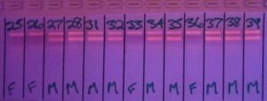
The DNA of the embryo needs to be multiplied to have the mass required to see it during the final phase of this sexing technology: gel electrophoresis. The content of each test tube containing the multiplied DNA is placed into a separate well in a prepared gel. An electric current is introduced across the gel, which causes all fragments of DNA to move with the current. The DNA fragments move according to electrical charge and fragment size. The male DNA fragments are smaller and thus separate from other (autosomal) DNA fragments. It takes approximately 20 minutes for the male DNA fragments to fully separate from the autosomal DNA, and a bright band of male DNA will show up when the gel is illuminated with ultraviolet light. By reading the specific positions of the bands of DNA, we can now tell which embryo is male and which embryo is female. The accuracy of this test is 98%.

The sex of embryo can then be selected for transfer.

AFTER THE EMBRYOS ARE SEXED

TRANSFER OR FREEZE

Historically all sexed embryos were transferred fresh to synchronized recipients. The demand for recipients is reduced when only the female embryos are transferred. For example, if 4 donors are flushed and 24 embryos are collected (average of 6 embryos per donor), approximately 12 synchronized recipients are required, as 50% of the embryos sexed should be female. The savings to the dairy or ranch in reducing the recipient pool maintenance costs can be substantial.

Sexed embryos are successfully frozen and thawed with a pregnancy rate that is equivalent to the pregnancy rate of unsexed frozen embryos.

THE ECONOMICS OF EMBRYO SEXING

Embryo sexing technology has remarkable accuracy, and can produce more female calves on the farm. It is also possible that embryo sexing can ultimately be less costly than a normal embryo transfer program alone.

Apart from the potential increases in genetic gain and marketability (of both sexed embryos and recipients carrying a fetus whose sex is known), embryo sexing enables producers to influence the overall cost of embryo transfer by reducing the required number of recipients.

Ultimately, total transfer costs are reduced. Historically, if more embryos were recovered than the number of available recipients, the extra embryos were simply frozen. To transfer these embryos at a later date requires synchronizing another group of recipients and the additional costs of transferring the frozen embryos. Assuming male embryos are of no value, sexing embryos reduces the total number of transferable embryos by half, thereby reducing the number of recipients that are used. This ensures that all recipients will be carrying embryos valuable to the genetic goals of the herd.

Some export markets are currently exploring the option to purchase only embryos of a desired sex (females for breeders, or males for A.I. units) to fill their orders; therefore, breeders using these technologies have a marketing advantage.

IVF TECHNOLOGIES

The Abbotsford Veterinary Clinic is one of only two facilities in Canada with an in vitro fertilization laboratory certified for international export of embryos.

Ovum pick-up and in vitro fertilization (OPU and IVF) are used primarily on infertile, pregnant, very young or very old donors. They are not recommended for donors with fertility problems having a genetic basis, but can be a very effective means of circumventing infertility that results from injury, disease or old age. In addition, the ability to obtain offspring from pregnant donors (40 – 100 days pregnant) and heifers as early as eight or nine months old can be very valuable to producers.

Donors can be aspirated as frequently as every two weeks, making it possible to obtain many calves from a particular donor in a short period of time from a selection of different sires.

Certain risks are involved with OPU such as ovarian adhesions and infertility particularly among very young donors, as well as the slight risk of abortion in pregnant donors. Anyone interested in OPU and IVF needs to be aware of these risks and work with our team to apply this technology appropriately.

THE TECHNOLOGY

Recently IVF technology has improved greatly and is now more widely commercially available.

In order to perform IVF, oocytes (unfertilized eggs) need to be collected from a superovulated donor through ovum pick-up. This is accomplished with use of an ultrasound to visualize the donor’s ovary and follicles. Oocytes are aspirated from these follicles using a sharp needle guided by the ultrasound probe. Extracted oocytes are incubated and continue to mature until they are ready for fertilization in the lab.

In vitro fertilization produces embryos via introduction of semen to mature oocytes. Once fertilized, embryos are cultured and evaluated at day 7, after which viable embryos are either frozen or transferred to recipients. Precise temperature controls are vital at all stages of handling oocytes, semen and embryos to ensure suitable embryo development.

THE ECONOMICS OF IVF

The cost per pregnancy for IVF embryos is generally higher than with embryo transfer, but it is a useful tool for obtaining offspring from valuable donors in situations where traditional breeding or embryo transfer would not be possible or productive.